Yeh **existing Diabetic Retinopathy page mein add karne wala HTML section** hai—poora page replace mat karna. Elementor ke **HTML widget** mein paste karein: diagnosis section ke baad, treatment section se pehle. Retina specialist se content review karwa kar hi live update karein.```html
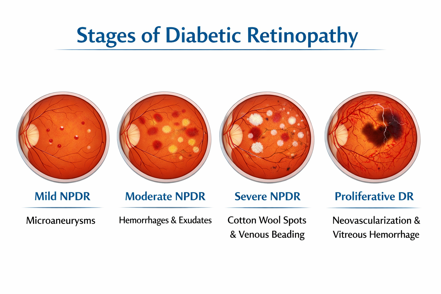
Understanding the Stages of Diabetic Retinopathy
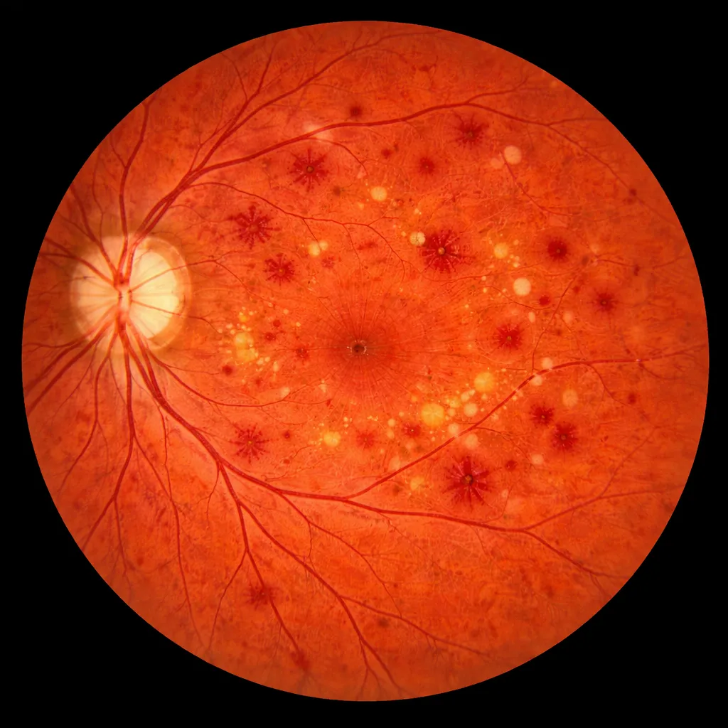
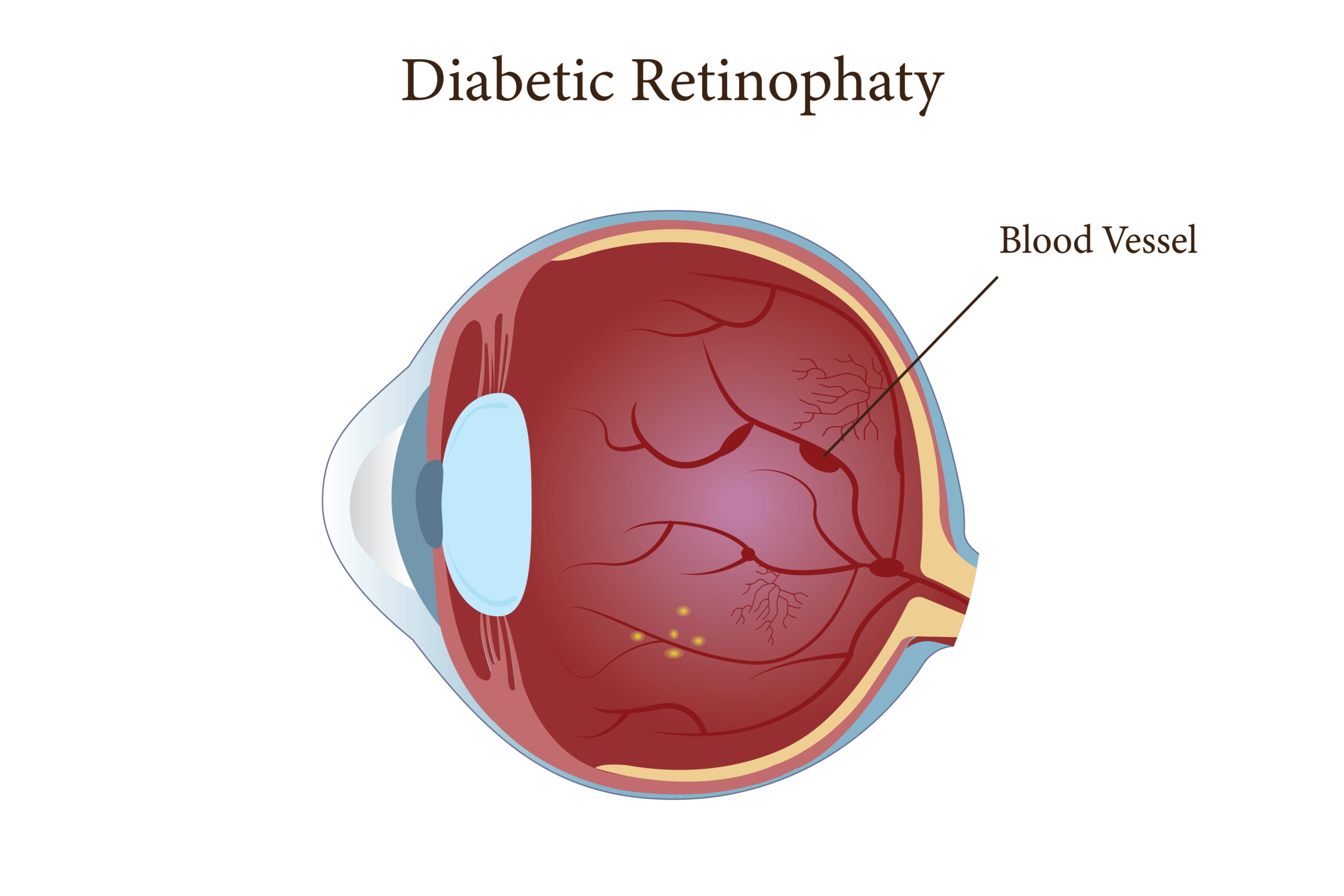
Diabetic retinopathy develops when diabetes affects the small blood vessels
in the retina. Early changes may not cause noticeable symptoms, so a person
can have retinal disease while still seeing clearly. An eye examination is
needed to identify and grade these changes.
Non-proliferative diabetic retinopathy (NPDR)
NPDR is described as mild, moderate or severe according to the retinal
findings. Small vessel changes, tiny areas of bleeding or reduced blood
flow may be seen. The recommended follow-up depends on the stage and
individual risk factors.
Proliferative diabetic retinopathy (PDR)
In PDR, abnormal new blood vessels can grow on the retina or optic nerve.
These vessels may bleed into the vitreous or contribute to scar tissue
and traction on the retina. A retina specialist should assess suspected
PDR promptly.
Diabetic macular edema can occur at different stages.
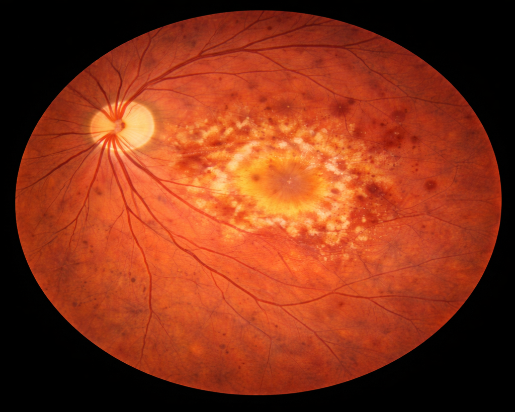
DME is swelling in the macula, the central part of the retina used for
detailed vision. It is assessed separately because it can affect central
vision and may change the treatment plan.
Learn about diabetic macular edema
.
Tests Your Retina Specialist May Recommend
A dilated retinal examination is used to look for diabetic retinal changes.
Retinal photographs may help document findings. Optical coherence tomography
(OCT) creates cross-sectional images of the retina and is commonly used when
macular swelling is suspected or needs monitoring.
Fluorescein angiography (FFA) uses injected dye and retinal photographs to
show blood flow and leakage. It is not required for every patient; a
specialist may recommend it when more detail about retinal circulation is
needed, including in selected cases of more advanced disease or DME.
How Treatment Decisions Are Made
Treatment is based on the retinal examination, stage of disease, presence
and location of DME, vision, other health factors and follow-up needs. The
table gives general examples only; it is not a treatment recommendation for
an individual.
| Finding | What the care plan may include |
|---|
| No or early retinopathy, without DME |
Scheduled eye examinations and monitoring. The clinician may also
discuss blood sugar, blood pressure and cholesterol management with
the patient’s diabetes-care team. |
| Moderate or severe NPDR |
Closer retinal follow-up. A retina specialist decides whether
monitoring or treatment is appropriate based on the individual
findings and risk of progression. |
| PDR |
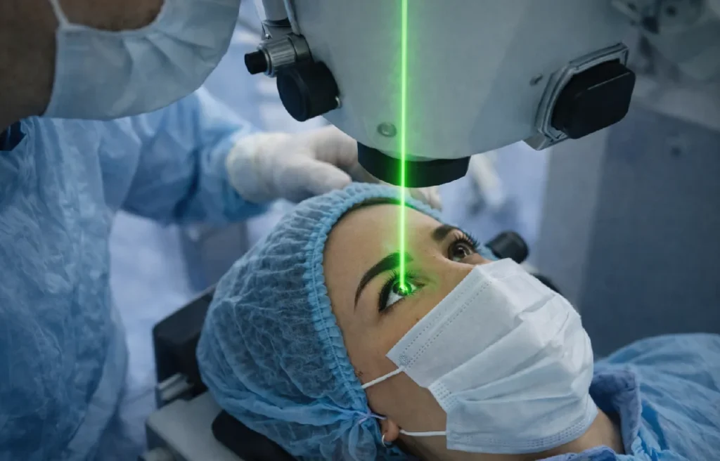
Treatment may include panretinal laser, intravitreal anti-VEGF
injections, or another approach selected by the specialist. Keeping
scheduled follow-up is important. |
| DME affecting the macula |
OCT-guided monitoring and, when indicated, intravitreal injections
or other treatments such as laser or steroid therapy may be
considered by the retina specialist. |
| Vitreous bleeding or tractional complications |
The specialist assesses the cause and severity. Vitrectomy may be
considered in selected advanced cases. |
How Often Should You Have a Retina Check?
Do not wait for blurred vision before arranging diabetic eye screening.
Many people with diabetes need a dilated eye examination at least once a
year, while people with retinopathy or DME may need more frequent reviews.
Your eye specialist should set the interval for your individual findings.
If you have pre-existing type 1 or type 2 diabetes and are planning a
pregnancy or are pregnant, tell your diabetes-care team and eye specialist
early. Retinopathy may need closer monitoring during this period. Do not
change diabetes medicines without advice from your treating doctor.
Book a Retina Evaluation
Medical References
This information is general education and does not replace an eye
examination or advice from your clinician.